Home » Without Label » 27+ schön Foto Picture Of The Inner Ear : Inner Ear Wikipedia / Let's start with a picture of the whole inner ear, which is the part of the ear that contains the cochlea.
27+ schön Foto Picture Of The Inner Ear : Inner Ear Wikipedia / Let's start with a picture of the whole inner ear, which is the part of the ear that contains the cochlea.
27+ schön Foto Picture Of The Inner Ear : Inner Ear Wikipedia / Let's start with a picture of the whole inner ear, which is the part of the ear that contains the cochlea.. The following picture shows a diagram of the right ear as it appears if you are looking at the cat's head from the front. Ménière's disease is a disorder of the inner ear that causes severe dizziness (vertigo), ringing in the ears (tinnitus), hearing loss, and a feeling of fullness or congestion in the ear. Your middle ear has bone linking the eardrum to the inner ear. Below are the six most common dog ear problems with pictures, and how to prevent and fix them. But, if you ignore it, then you can suffer from permanent hearing loss.
The outer ear, the middle ear and the inner ear. One of the most often encountered dog ear problems is associated with ear. It is also the site of the opening to the ear canal. The eardrum is responsible for separating the middle and outer ear. The inner ear is at the end of the ear tubes.
Otitis Externa Ear Canal Outer Ear Inner Ear Ear People Abroad Sound Png Pngwing from w7.pngwing.com There are three components to the ear: The inner ear is at the end of the ear tubes. Your doctor can diagnose it based on your pattern of symptoms and a medical evaluation. They are the outer ear (the part we see along the sides of our head behind the temples), the middle ear, and the inner ear.but in terms of function, the ear has four parts: The inner ear has 3 main parts: Browse 204 ear canal stock photos and images available, or search for inner ear or hearing loss to find more great stock photos and pictures. See human ear anatomy stock video clips. When they are dislodged, the crystals float around in the fluid area of the balance branch of the inner ear, and you will start to feel off balance.
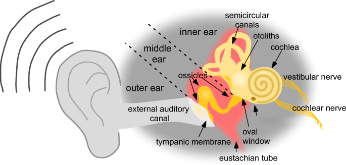
Illustration of hearing, journey of the sound wave in the ear.
Your middle ear has bone linking the eardrum to the inner ear. The ear anatomy and function of the ear. The sound wave is captured by the auricle, penetrates auditory canal, vibrates eardrum. Your doctor can diagnose it based on your pattern of symptoms and a medical evaluation. Within these cells are tiny particles (otoconia) that help monitor the position of your head in relation to. Illustration of hearing, journey of the sound wave in the ear. There are three components to the ear: The ear is composed of the outer ear, middle ear, and inner ear. Below are the six most common dog ear problems with pictures, and how to prevent and fix them. Browse 1,523 inner ear stock photos and images available or search for inner ear illustration or inner ear diagram to find more great stock photos and pictures. The inner ear is at the end of the ear tubes. The inner ear connects to the brain and contains nerves and centers for balance and hearing. You don't need expensive tests to get a diagnosis of bppv.
Seventeen year old male with a two day history of ear pain and sore throat. See human ear anatomy stock video clips. The outer ear, the middle ear and the inner ear. Human ear the ear is the organ that detects sound. Attacks of dizziness may come on suddenly or after a short period of tinnitus or muffled hearing.
Antisense Oligonucleotides For The Treatment Of Inner Ear Dysfunction Springerlink from media.springernature.com The sound wave is captured by the auricle, penetrates auditory canal, vibrates eardrum. Red dilated blood vessels at the upper part of the ear drum. You can see it in the picture at the top of this page. The ear anatomy is divided into three parts, the outer, middle, and inner ear. The eardrum is responsible for separating the middle and outer ear. Each section performs distinct functions that help transform vibrations into sound. Infection affecting the inner ear is referred to as otitis media. The infection can trigger temporary hearing loss.
The ear is part of the auditory system and is the organ that detects sound.
Just as with a myringotomy, the doctor will make an incision in the eardrum and suction out the fluid that's accumulated in the middle ear. Ear cancer can affect both the inner and external parts of the ear. Browse 1,523 inner ear stock photos and images available or search for inner ear illustration or inner ear diagram to find more great stock photos and pictures. See inner ear stock video clips. Illustration of hearing, journey of the sound wave in the ear. If you are 60 or older, you are more prone to having your ear crystals dislodge. Labyrinthitis (vestibular neuritis) is the inflammation of inner ear. At the base of the canals are the utricle and saccule, each containing a patch of sensory hair cells. Your doctor can diagnose it based on your pattern of symptoms and a medical evaluation. Ménière's disease usually affects only one ear. The inner ear consists of tiny bony structures filled with fluid. Six year old with an early ear infection. It often starts as a skin cancer on the outer ear that then spreads throughout the various ear structures, including.
Infection affecting the inner ear is referred to as otitis media. The inner ear consists of tiny bony structures filled with fluid. The loose crystals will start to make people feel like they are spinning and the room is spinning around them. As sound waves travel from the outer to the inner ear, they create waves in the fluid of the inner ear, which in turn moves the tiny hairs in the ear that send sound or movement. There are three sections of the ear, according to the anatomy textbooks.
Inner Ear Anatomy And Barriers A A Schematic Drawing Of The Download Scientific Diagram from www.researchgate.net Cats do not have as many ear problems as do dogs. If you are 60 or older, you are more prone to having your ear crystals dislodge. Let's start with a picture of the whole inner ear, which is the part of the ear that contains the cochlea. There are three components to the ear: The ear anatomy of ear ear anatomy the human ear anatomy of the ear ear diagram ear structure diagram of ear inner middle ear human ear parts. When the inner ear is inflamed or irritated, symptoms such as dizziness, loss of balance, ringing in the ear , nausea, and vomiting may come on suddenly. The auditory ossicles of the middle ear and the structures surrounding them. Below are the six most common dog ear problems with pictures, and how to prevent and fix them.
The ear anatomy and function of the ear.
There are three components to the ear: The inner ear consists of tiny bony structures filled with fluid. You can see it in the picture at the top of this page. When they are dislodged, the crystals float around in the fluid area of the balance branch of the inner ear, and you will start to feel off balance. The sound wave is captured by the auricle, penetrates in the auditory canal, vibrates. See inner ear stock video clips. Ménière's disease is a disorder of the inner ear that causes severe dizziness (vertigo), ringing in the ears (tinnitus), hearing loss, and a feeling of fullness or congestion in the ear. Less commonly, an inner ear infection is a true infection caused by a virus or bacteria. The loose crystals will start to make people feel like they are spinning and the room is spinning around them. At this point, he will insert a small tube into the ear drum to ventilate the middle ear. The auditory ossicles of the middle ear and the structures surrounding them. Ménière's disease usually affects only one ear. Labyrinthitis (vestibular neuritis) is the inflammation of inner ear.